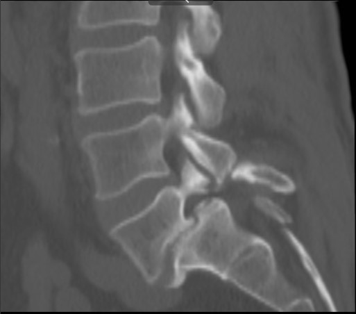
RADIOLOGICAL ASSESSMENT OF SPONDYLOLISTHESIS OF LUMBOSACRAL SPINE
DOI:
https://doi.org/10.48026/issn.26373297.2024.1.15.3Keywords:
SPONDYLOLISTHESIS, MRI of the spine, CT of the spine, lumbosacral spine, radiology imaging of the spineAbstract
Introduction: One major injury or frequent minor injuries cause the vertebral body, pedicle and superior articular surfaces to slide forward. This disorder is known as spondylolisthesis and is usually accompanied by symptoms, often proportional to the degree of forward movement. Patients may complain of pain in the lower back that spreads to the thighs, and limited range of motion is possible
Objectives: The aim of this study is to determine the frequency of spondylolisthesis by age and gender in the patients studied at SKB Mostar.
Research methodology: The method of content analysis was used to retrospectively examine X-ray, CT and MRI examinations of patients with the clinical features of lumbar pain syndrome in the period from 1 January 2019 to 1 January 2020 in patients who underwent radiological examination of the LS spine at the Clinical Institute of Radiology of the University Clinical Hospital Mostar. Data were collected from IMPAX at the SKB Mostar Department of Radiology. The input parameters that were observed were: pain syndrome, diagnosis. The output parameters were the radiological findings from the X-ray, CT, and MRI scans.
Results: During the study period, 297 spinal scans were performed, and spondylolisthesis was found in 32 patients, or 11%. The youngest respondent was 8 years old, and the oldest was 85 years old. The average age of the respondents was 64.37 years. The largest number of respondents was in the age group of over 75 years old. Of the tests performed, the most common were CT scans, followed by MRI, and the rarest were X-ray scans. The highest prevalence of spondylolisthesis was found in vertebrae L4-L5 and L5-S1 at 41%. Spondylolisthesis was found bilaterally in 9% of cases at the L3-L4 and L5 vertebrae.
Conclusion: Most often, spondylolisthesis is found in L4-L5 and L5-S1 vertebrae. There was no difference according to gender, and most often subjects older than 75 years had spondylolisthesis.

Downloads
Published
How to Cite
Issue
Section
License
Copyright (c) 2024 Administrator administrator

This work is licensed under a Creative Commons Attribution 4.0 International License.
Copyright & licensing:
This journal provides immediate open access to its content under the Creative Commons CC BY 4.0 license. Authors who publish with this journal retain all copyrights and agree to the terms of the above-mentioned CC license.


